MGD is a chronic and progressive condition1,2,3
The outermost layer of the tear film, the lipid layer, is composed of oils from meibomian gland secretions that lubricate, prevent evaporation, and perform barrier functions. If glands become obstructed, qualitative and quantitative changes in glandular secretion may lead to symptoms of eye irritation, clinically apparent inflammation, and ocular surface disease.4
In one study, up to 86% of dry eye patients had signs of MGD.5
Understanding MGD
View the videos below to learn about the role of meibomian glands in ocular health
Ocular surface and gland function in a healthy eye
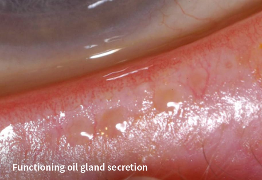
In a healthy eye, pressure from a blink expresses a small amount of oil from the meibomian glands which is then distributed over the ocular surface as the eye opens. The ocular surface is the foundation for ocular comfort and visual quality.6
Impact of blocked meibomian glands
Obstruction of meibomian glands impedes the production of oils necessary to reduce aqueous evaporation and minimize harmful friction between the eyelids and cornea.
Long-term effects of untreated MGD
If left untreated, obstructed glands will reduce oil production, atrophy, and eventually drop out. Once a gland has atrophied completely, function is lost permanently, which leads to chronic discomfort and potentially sight-threatening damage to the ocular surface.
Functioning vs. Blocked Glands

MGD and Dry Eye Facts
 |
In one study, up to 86% of dry eye patients had signs of MGD5 |
 |
Over 745 million people worldwide suffer from dry eye8 |
 |
In one study, 63% of cataract patients have an unstable tear film7 |
 |
In one study, 60% of contact lens wearers had a sign of MGD10 |
The ocular surface is the foundation for ocular comfort and visual quality and it is impaired in much of the general population4,6
Assessing Meibomian Glands
MGD CAN BE EVALUATED BASED ON OBSERVABLE COMPROMISE TO GLAND FUNCTION AND STRUCTURE.9
Function
By simulating the pressure of a deliberate blink, the Meibomian Gland Evaluator (MGE) provides a standardized, repeatable evaluation of meibomian gland function during slit lamp examinations.
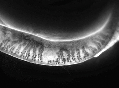
Structure
Dynamic Meibomian Imaging (DMI) technology available with LipiScan® Dynamic Meibomian Imager and LipiView® II Interferometer simultaneously employs surface illumination and high-definition transillumination to provide an accurate visualization of gland morphology.
Improving Gland Function

The unique technology of the LipiFlow® Thermal Pulsation System has been shown to improve gland function in patients with MGD.10
By simultaneous application of heat and peristaltic motion to the eyelid, obstructed meibum is safely liquefied and pushed out of the gland orifices.
Images for illustrative purposes only. Actual results may vary. Not actual patients or healthcare professionals.